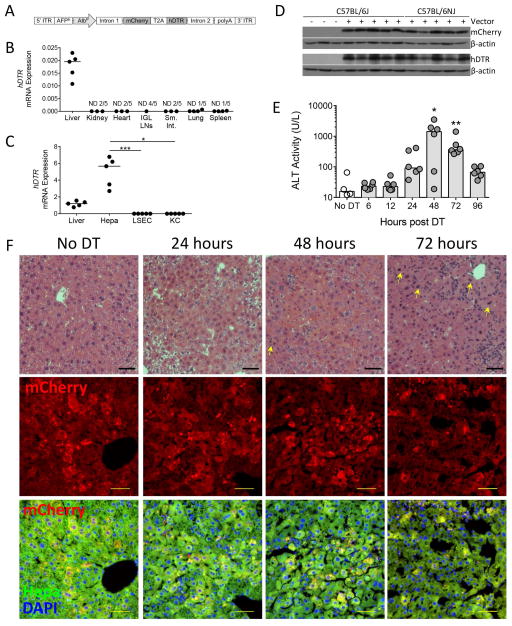
Figure 1. Hepatocyte-specific expression of rAAV8.pAlb.mCherry.hDTR results in peak liver damage 48 hours following DT injection.
(A) rAAV8.pAlb.mCherry.hDTR vector encoding the mouse AFPE and AlbP; mCherry and the hDTR linked by a T2A linker sequence; and AAV serotype-2 5′ and 3′ ITRs. Two weeks following rAAV injection, mice received either no injection, 1X PBS (control), or DT and were sacrificed 6, 12, 24, 48, 72, or 96 hours later. hDTR mRNA levels, relative to (B) Gapdh in organs and (C) Hprt in total liver and liver cell populations of C57BL/6NJ mice that received 1X PBS, each data point represents an individual mouse (n = 5). (D) Western blot detection of mCherry, hDTR, and β-actin in the hepatocytes of C57BL/6J mice that did not receive rAAV and in C57BL/6J and C57BL/6NJ mice that received rAAV and 1X PBS. (E) Serum ALT levels in C57BL/6J mice that received no DT or DT. Each data point represents an individual mouse, bars represent the median. (F, top row) Paraffin-embedded sections stained for hematoxylin and eosin, 100X. Bar, 50 μm. Arrow, mitotic hepatocytes. (F, middle and bottom rows) Cryo-preserved sections with mCherry (top) or the merge of mCherry fluorescence (red), hepatocyte autofluorescence (green), and DAPI to distinguish nuclei (blue) (bottom). Bar, 50 μm. (D–F) are combined from two experiments (n = 4–6 per group). Representative images chosen from one mouse at each time point for (F), based on median ALT levels. Significance determined by a Kruskal-Wallis test followed by Dunn’s post-test (C) of Liver, LSEC, and KC to hepatocytes and (E) of each time point to the No DT group median. * represents significance compared to No DT group; *, P ≤ 0.05; **, P ≤ 0.01; ***, P ≤ 0.001; non-significance represented by absence of bar or asterisk. AFPE, AFP enhancer; AlbP, albumin promoter; DAPI, 4′,6-diamidino-2-phenylindole; DT, diphtheria toxin; Hepa; hepatocyte; hDTR, human diphtheria toxin receptor; ITR, inverted terminal repeats; IGL, inguinal lymph nodes; KC, Kupffer cell; LSEC, liver sinusoidal endothelial cell; Sm. Int., small intestine; and ND, non-detect.

