FIGURE 5:

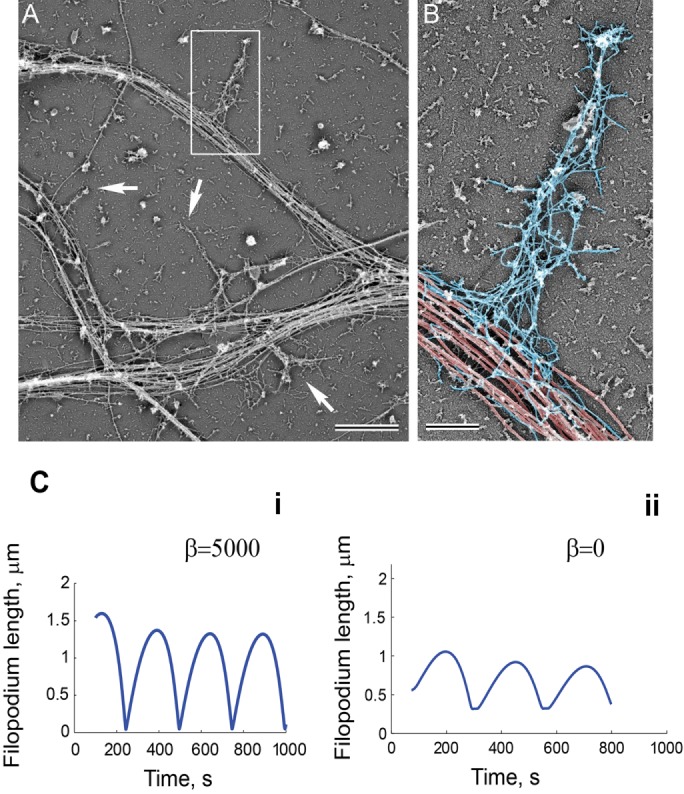
Filopodia dynamics depends on the resistive force applied at the base by the microtubule network. Cytoskeletal organization of dendritic filopodia revealed by platinum replica electron microscopy. (A) A network of dendrites from hippocampal neurons cultured for 8 DIV. Dendritic filopodia (boxed region and arrows) reside on dense arrays of microtubules in dendrites. (B) Dendritic filopodium from the boxed region in A color-coded to show microtubules in the dendrite (red) and actin filaments in the filopodium (blue). Scale bars, 2 μm (A), 0.5 μm (B). (C) Simulated filopodia dynamics with (i) and without (ii) resistive force at the barrier. (i) L0 = 1, koff = 0.13, kon = 0.12, vp = 0.9, m0 = 25, η = 100, ζ = 100, σ0 = 25, β = 5250, and D = 0.04. (ii) L0 = 1, koff = 0.13, kon = 0.12, vp = 0.9, m0 = 25, η = 100, ζ = 100, σ0 = 25, β = 0, and D = 0.04.
