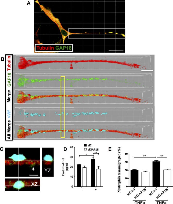
FIGURE 6:
ARHGAP18 colocalizes with vWF+ WPBs and contributes to regulation of cargo release. (A) EC stained with ARHGAP18 (green) and tubulin antibodies (red) and imaged with confocal microscopy (630× magnification). White box indicates region zoomed in B. (B) Extended process of an EC; tubulin staining (Alexa 647; red), ARHGAP18 (Alexa 488; green), and ARHGAP18 puncta shown along MTs in Merge. Images are rendered in Imaris software (shadow projection mode). vWF immunopositivity is shown in cyan (Alexa 594). All Merge, ARHGAP18, tubulin, and vWF channels merged together. Yellow box indicates region of interest zoomed in C to show colocalization of ARHGAP18 (green) with vWF (cyan) in a WPB attached to the MT filament. Smaller ARHGAP18 puncta (as observed in GSD imaging) are seen to the right of the WPB (white arrowhead). XZ- and YZ-projections at the cross-hair point indicate colocalization of immunopositivity and the WPB localization to the MT. Scale bar, 30 μm (A), 10 μm (B), 1.5 μm (C). (D) ECs were transfected with control siRNA (siC; ■) or siRNA for ARHGAP18 (siGAP18; □) and were stimulated after 48 h with PMA (+) for 30 min. The supernatant was collected and analyzed for expression of soluble endothelin-1 by ELISA. Results are mean ± SEM of four individual HUVEC lines, *p < 0.05, **p < 0.01. (E) ECs were transfected with control siRNA (siControl; □) or siRNA for ARHGAP18 (siGAP18; ■) and were stimulated after 72 h with TNFα for 4 h. Neutrophils were allowed to transmigrate through ECs for 1 h and then collected; the number of neutrophils was determined and is expressed as a percentage. Results are mean ± SEM of triplicate determinations of each group from one experiment representative of three experiments performed; **p < 0.01.

