
FIGURE 8:
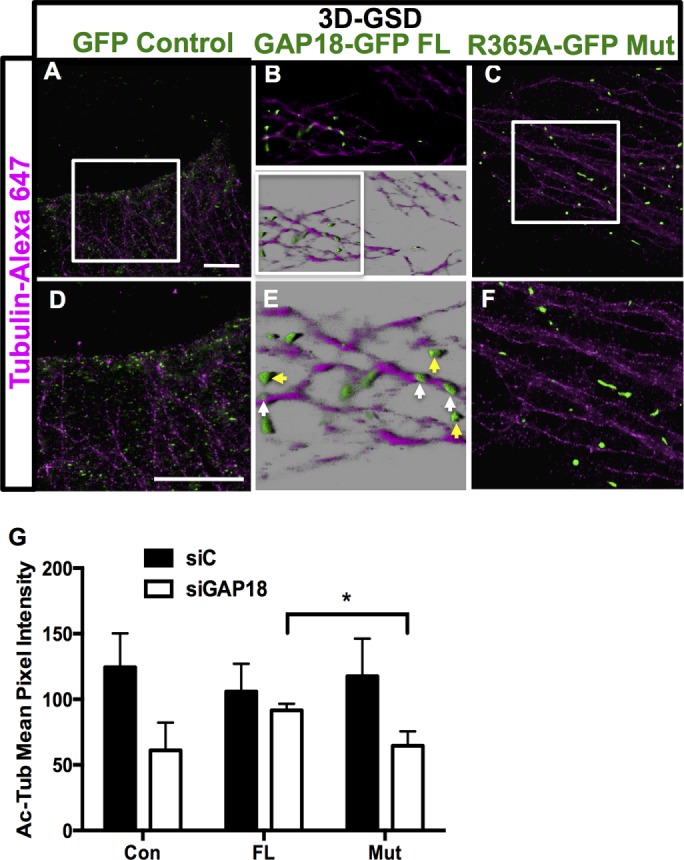
A functional RhoGAP domain in ARHGAP18 is required for MT localization and stability. GFP-ARHGAP18 or GFP-control plasmid was transfected into HeLa cells. (A) The 3D GSD of control plasmid; zoom of the white box is shown in D. (B) The 3D GSD imaging of full-length, wild-type ARHGAP18-GFP puncta in the fringe of a HeLa cell. Maximum intensity projection (top) and 3D reconstruction in Imaris software blend mode (bottom). (E) Zoom of white box in B. Individual ARHGAP18 puncta elongated along MTs are indicated by white arrows; cytosolic puncta are shown in yellow. (C) Lack of localization of ARHGAP18 R365A mutant constructs to MTs is observed. (F) Zoom of white box in C. Scale bar, 5 μm (A, D). (G) Rescue of the disrupted MT phenotype by functional ARHGAP18. ECs were transfected with siRNAs for control (■) or siARHGAP18 (□). At 24 h later, they were transfected with either GFP-control plasmids (Con), wild-type full-length GFP-ARHGAP18 (FL), or GFP-mutant ARHGAP18 (Mut). Eighteen hours later, cells were fixed and stained for Ac-Tub. Cells that were GFP positive, indicating transfection with the plasmids, were analyzed for the extent of Ac-Tub area and pixel intensity. Results from 60 GFP-positive cells from three separate experiments; *p < 0.05.