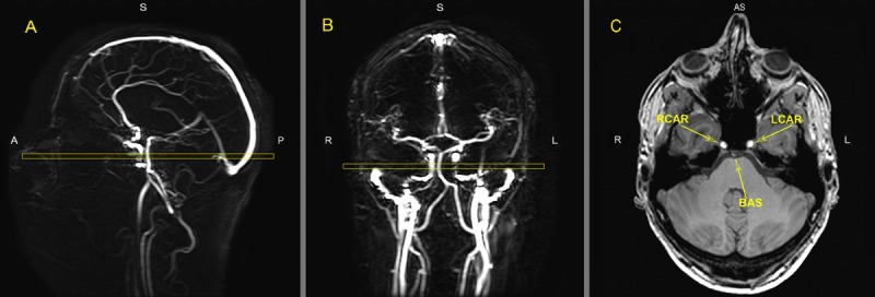
Figure 5. MR images of the vasculature measured with phase contrast MR angiography imaging for Quantitative Flow (QF).

Typical placement of the QF phase-contrast slice (C) through the right internal carotid artery (RCAR), left internal carotid artery (LCAR) and the basilar artery (BAS) using the sagittal (A) and coronal (B) localizer angiograms. ROI's were drawn around the three arteries in slice C to measure flow.
