
Fig. 2.
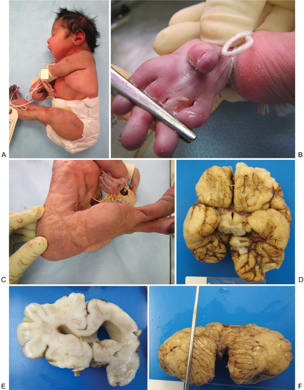
Neonatal phenotype and pathology. (A) Lateral view of newborn demise, showing microcephaly, arthrogryposis, and collapsed auricular tragus. (B) Close-up view of the right hand showing elongated fingers, shortened thumb, and absent palmar creases. (C) The lateral view is showing arthrogryposis, decreased musculature, shortened forelegs, and redundant dorsal foot skin. (D) Basilar view of the neonatal brain at autopsy showing asymmetry, with smaller right cerebrum and disruption of cerebral cortical development with agyria/pachygyria of bilateral temporal and right frontoparietal lobes. (E) Coronal section of the neonatal brain showing asymmetric ventriculomegaly. (F) Dorsal view of the neonatal cerebellum showing Dandy–Walker malformation with near-complete agenesis of the vermis.