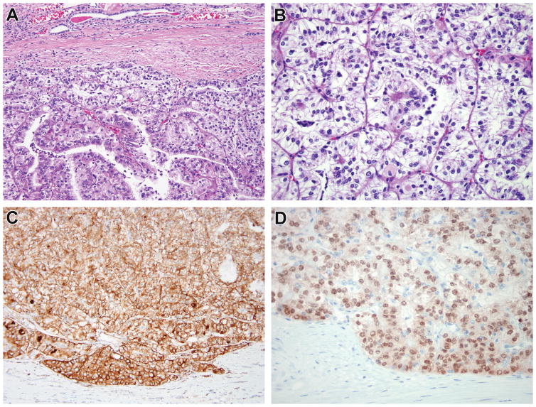
Figure 3.
Case 3. This renal cell carcinoma demonstrates nested and papillary architecture and epithelioid cells with clear to eosinophilic cytoplasm (A, B). A subpopulation of smaller cells is present within the lumens of acini. The neoplastic cells are diffusely immunoreactive for cathepsin k (C). While conventional TFE3 FISH did not demonstrate rearrangement, the neoplasm demonstrated diffuse, strong nuclear labeling for TFE3 protein with a clean background (D), prompting additional FISH fusion studies which demonstrated the RBM10-TFE3 gene fusion.

