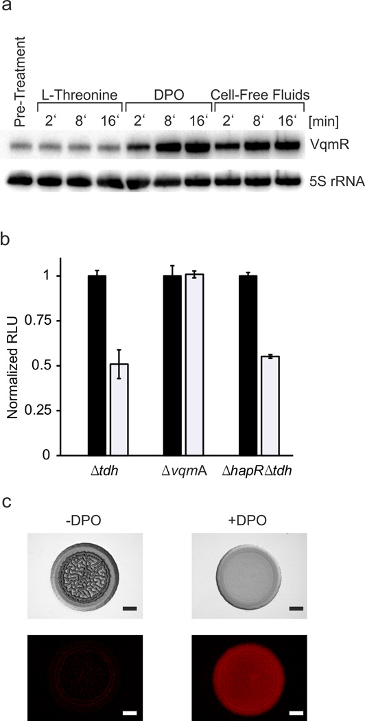
Fig. 5. DPO induces production of VqmR which represses V. cholerae biofilm formation.
(A) Northern blot showing VqmR production in the V. cholerae Δtdh strain grown in M9 medium with casamino acids and collected at the designated time points following addition of 1 mM L-threonine, 100 µM DPO, or cell-free culture fluids prepared from wild-type V. cholerae (25% final conc.). The first lane shows the no addition control. 5S rRNA served as loading control. Source files with blots showing the full data can be found in Supplementary Fig. 8D. (B) The designated V. cholerae mutants carrying a PvpsL::lux reporter fusion were supplied without (black bars) or with (white bars) 100 µM DPO. Bioluminescence emission and OD600 were measured at 5.5 h to obtain relative light units (RLU). In each set, the RLU of the untreated sample was set to 1.0 for normalization. Each experiment was performed in triplicate. Error bars correspond to one standard deviation from the mean. (C) A ΔhapR, tdh::Tn5, pvqmR::mKate2 V. cholerae strain harboring PvqmR on a low copy plasmid was grown in liquid LB medium with chloramphenicol to OD600 = 0.7 prior to being spotted onto an LB agar plate lacking or containing 1 mM DPO. Transmission and fluorescence (mKate2) images were captured after 2 days of growth at 30 °C. The experiment was performed in duplicate 10 times and a representative image is shown for each condition. Scale bar = 1 mm.

