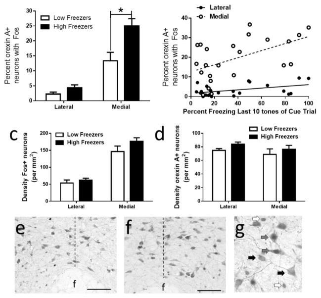
Figure 2.
Activation of orexin neurons in the lateral and medial regions of the hypothalamus in high and low freezers. (a) High freezers with poor extinction recall had a significantly greater percentage of orexin A positive neurons expressing Fos in the medial hypothalamus than low freezers. (b) Mean percent freezing during the last ten tone presentations of within trial extinction was positively correlated with the density of dual labelled neurons in both the lateral and medial regions of the hypothalamus. No overall differences in Fos positive neuron density (c) or orexin A positive neuron density (d) between high and low freezers were observed. Representative photomicrographs showing Fos and orexin A immunoreactivity in the perifornical region of the hypothalamus in low (e) and high (f) freezers (20X; scale bar=100 microns) and high magnification (40X) of Fos positive (white arrows), orexin A positive (black arrows) and dual labelled neurons (gray arrows) in the medial hypothalamus. Dotted line represents the level of the fornix, separating medial and lateral hypothalamus. * indicates P<0.05.

