Fig. 6.
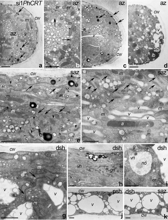
Ultrastructure of Petunia si1PhCRT pollen tubes. a–d The apical zone (az) contains numerous mitochondria (m), very short or even fragmentized/disorganized ER (er and arrows in b), electron-dense vesicles (arrows in a, c), vacuoles (v), and lipid bodies (lb). e, f In the subapical zone (saz), only single ER cisternae (arrows in e) are present; mitochondria (m) and dictyosomes (arrowheads in e) are normally localized in saz; however, dictyosomes are observed mainly in the peripheral cytoplasm (arrows in f). g-i The distal shank (dsh) is highly vacuolated and occasionally contains ER (arrows in g and er in i), two-phase vesicles with electron-dense cortices (arrow in h), and normally positioned MGU (h) composed of the vegetative and generative nuclei (vn or gn). j The proximal shank (psh) is highly vacuolated. a amyloplast, cw cell wall. Bars 1 μm

