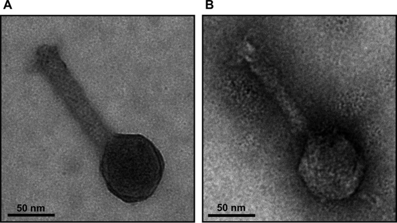
FIG 3.

Electron micrographs of isolated ϕCh1 particles. Virions resulting from lysis of the wild-type strain N. magadii L11 (A) and the ORF79 disruption strain N. magadii L11-ΔORF79 (B) were isolated and negatively stained with uranyl formate for electron microscopy.
