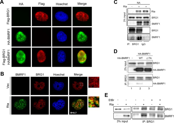
FIG 6.
BMRF1 associates with BRG1 in Rta-reactivated EBV-positive NA cells. (A) The Flag-BRG1-expressing plasmid was cotransfected with a vector control or HA-BMRF1 into HeLa cells for immunofluorescence analysis. The distributions of BRG1 and BMRF1 were detected by mouse anti-Flag and rabbit anti-HA antibody, respectively. Representative images of two independent experiments are shown. (B) At 48 h postinduction, slide-cultured NA cells were harvested for immunofluorescence analysis using mouse anti-BMRF1 and rabbit anti-BRG1 antibody. The distributions of BMRF1 and BRG1 were observed by confocal microscopy. Quantification of colocalization was measured by Pearson's correlation coefficient (r) using ImageJ Coloc2. Representative images of two independent experiments are shown. (C) NA cells transfected with a vector control or Rta expression plasmid were harvested for immunoprecipitation assays. Representative data of two independent experiments are shown. (D) The vector control or an HA-BMRF1 wild-type or transactivation domain mutant (ΔTA) plasmid was transfected into 293T cells for 48 h. The transfected cells lysates were harvested and subjected to immunoprecipitation assay using mouse anti-BRG1 antibody. The representative data of two independent experiments are shown. (E) Cell lysates harvested from control vector or Rta transfected NA cells were pretreated with or without EtBr (10 μg/ml) on ice for 30 min. Cell lysates were incubated with anti-BRG1 or control IgG to precipitate BRG1-associated complexes and subjected to Western blotting.

