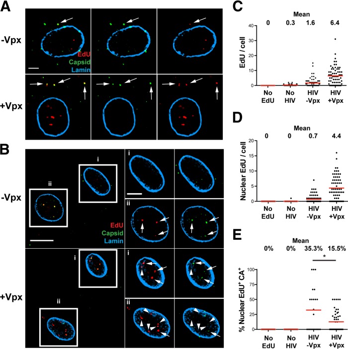
FIG 1.
Incorporation of EdU into HIV-1 particles in infected MDM. MDM were cultured without SIV-VLP (−Vpx) or with SIV-VLP (+Vpx) for 16 h and subsequently infected with VSV-G-pseudotyped HIVLAI∂env (HIV) in the presence of 10 μM EdU. (A and B) Cells were fixed after 24 h, labeled for EdU, stained for HIV-1 CA and nuclear lamin A/C, and imaged. (A) Representative z-sections of individual cells. Arrows indicate colocalized CA and EdU. (B, left) Representative z-sections of cells within a single field. (Right) Boxed cells (i and ii) showing enlarged images the same single z-sections of specified nuclei. Arrows denote colocalized intranuclear EdU and CA, and arrowheads denote EdU not colocalized with CA. (C and D) Numbers of total (C) and nuclear (D) EdU puncta per cell were quantified by using Imaris software. Mean values are indicated above the plots. No EdU, SIV-VLP-treated, HIV-1-infected cultures without EdU; No HIV, SIV-VLP-treated, EdU-containing cultures without HIV-1. (E) Percentage of EdU colocalized with CA in the nuclei of infected MDM. Fifty to seventy-five cells were analyzed under each condition in panels C to E. Data are representative of results from at least 3 independent experiments. A Mann-Whitney test was used to test for statistical significance in panel E (*, P < 0.05). Bars, 2.5 μm (A), 10 μm (B, left), and 5 μm (B, right).

