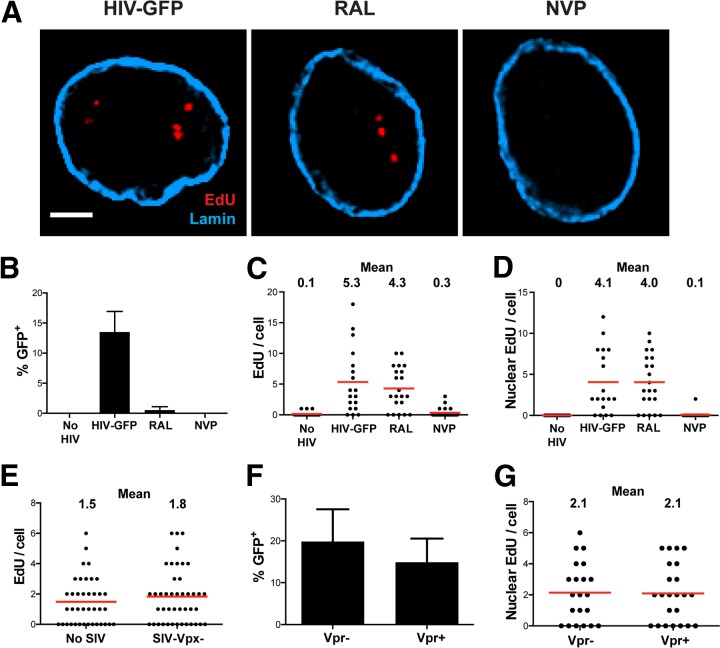
FIG 2.
EdU incorporation is dependent upon RT but not integration and is not the result of Vpr or SIV-VLP. MDM were cultured with SIV-VLP for 16 h and subsequently infected with VSV-G-pseudotyped HIVLAI∂env that expresses GFP in place of the nef gene (HIV-GFP). (A) MDM were washed at 16 h p.i., and cells were labeled for EdU, stained for lamin A/C, and imaged at 48 h p.i. (A) Representative z-sections of nuclei with no drug (HIV-GFP), 10 μM RAL, or 1 μM NVP. (B) Percentage of GFP+ cells (n = ∼200 under each condition). (C and D) Numbers of total (C) and nuclear (D) EdU puncta per cell quantified by using Imaris software (n = ∼30 cells under each condition). Graphs in panels B to D are representative of results from 3 independent experiments. (E) MDM were cultured for 16 h without (No SIV) or with (SIV-Vpx−) SIV-VLP containing a null vpx gene. MDM were infected and imaged as described above for panel A, and the total number of EdU puncta per cell was quantified. (F and G) MDM were cultured for 16 h with SIV-VLP containing a null vpr gene, infected with HIV-GFP containing a functional (Vpr+) or null (Vpr−) vpr gene in the presence of EdU, washed at 16 h, and cultured for a total of 4 days. Cells were quantitatively imaged for the percentage of GFP+ cells (F) and the number of intranuclear EdU puncta per cell (G). Bar, 2.5 μm (A). Error bars represent standard errors of the means from 3 random fields in panels B and F.

