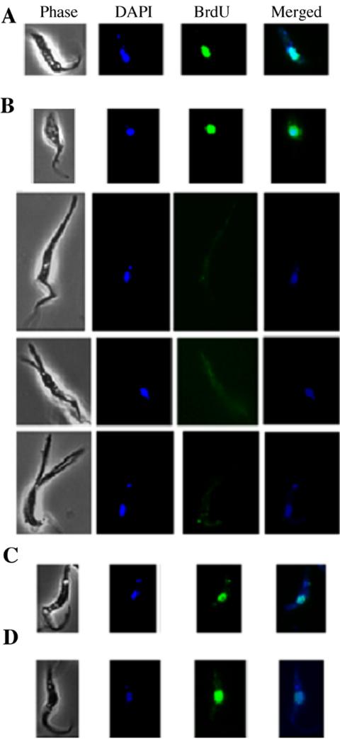
Figure 4.
BrdU incorporation in control and RNAi-treated cells. BrdU was added to strain 29-13 cells 3 d after RNAi induction, and the cells were harvested 2 d later. Images from the anti-BrdU immunofluorescence assays were recorded and superimposed on the DAPI staining patterns. All cells are oriented with their posterior ends pointing upward. (A) A control cell. (B) CRK1+CRK2-deficient cells. (C) A CRK1+CRK4-deficient cell. (D) A CRK1+CRK6-deficient cell. Note that BrdU only failed to incorporate into the nuclei of cells with elongated posterior ends.

