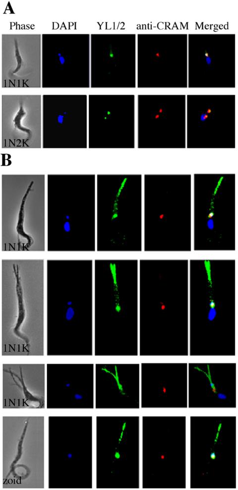
Figure 5.
Localization of newly assembled microtubules in CRK1+CRK2-depleted cells of abnormal morphology. Strain 29-13 cells after 5 d of CRK1+CRK2 RNAi were stained with DAPI, YL1/2 for tyrosinated α-tubulin, and anti-CRAM for the flagellar pocket and examined by fluorescence microscopy. All cells are oriented with their posterior end pointing upward. (A) 1N1K and 1N2K control cells without RNAi induction. (B) CRK1+CRK2-deficient 1N1K cells and a zoid with elongated and/or multiple posterior ends.

