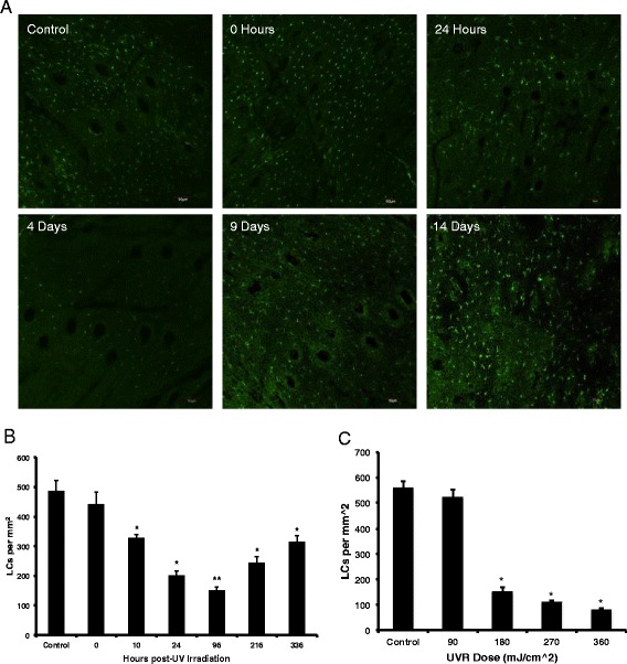
Fig. 5.

Langerhans cells (LC) migration kinetics. a The Langerhans cell (LC) migration was quantified by confocal microscopy of CD207 (langerin marker, Alexa 488-shown in green) stained hairless mouse epidermis measured from 0 h to 14 days after exposure to 180 mJ/cm2 UVB radiation, Scale Bar = 10 μm. b The bar chart represents the number of LCs per epidermal area over time quantified using ImageJ software. The graph represents the mean +/- standard error (SEM), N = 3, n = 3 (three regions analyzed for each epidermal sheet imaged). *p < 0.05, **p < 0.0001, 2-Talied t-Test, unpaired with unequal variances with respect to control. c LC migrations kinetics with respect to to UVR dose response at day 4 post-irradiation. *p < 0.05, 2-Tailed Students t-Test with unequal variance with respect to control
