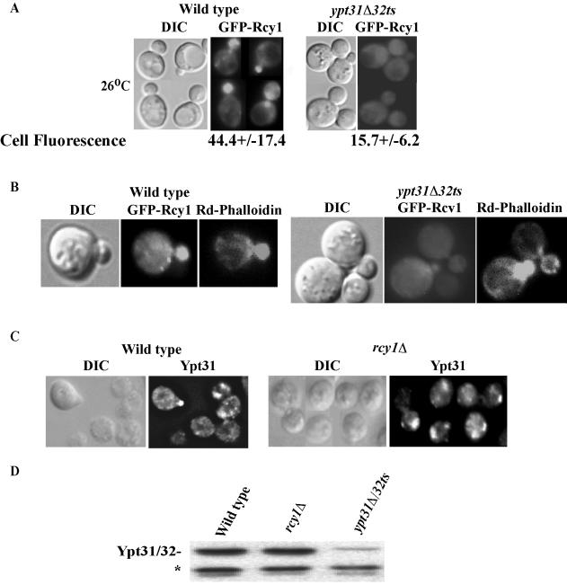
Figure 4.
Ypt31/32 affect the localization and the protein level of Rcy1 but not vice versa. (A) Ypt31/32 affect the localization of Rcy1. GFP-Rcy1 was expressed in wild-type (NSY125) and ypt31Δ/32ts mutant (NSY348) cells growing at 26°C. Cells were synchronized by incubation with α-factor (see Materials and Methods for details). GFP-Rcy1 protein localization was then examined by direct fluorescence microscopy. Bottom, cell fluorescence was quantified in Photoshop 7.0. Eleven random cells were quantified for each strain; ± represents the SEM. (B) Actin polarization is normal in ypt31Δ/32ts cells. Cells expressing GFP-Rcy1 were grown at 26°C, fixed, and stained with rhodamine-phalloidin, and then cells were examined by direct fluorescence microscopy. GFP-Rcy1 was viewed using an FITC filter and actin staining was viewed using a Texas Red filter. (C)Ypt31 localization is not disrupted in rcy1Δ mutants. Exponentially growing wild-type (NSY125) and rcy1Δ (NSY657) cells were fixed and Ypt31/32 localization was determined using affinity-purified anti-Ypt31p antibody and indirect immunofluorescence microscopy. In panels A–C, the contour of the cells is shown in the DIC image. (D) The level of Ypt31/32 is not affected by Rcy1. Protein level of Ypt31/32, in wild-type (NSY125), rcy1Δ (NSY657), and ypt31Δ/32ts (NSY348) cells was detected by Western blot analysis by using affinity-purified anti-Ypt31/32 antibody. Equal loading was confirmed by Ponceau S staining and a cross-reacting band that is present in ypt31Δ cells (*).

