Figure 6.
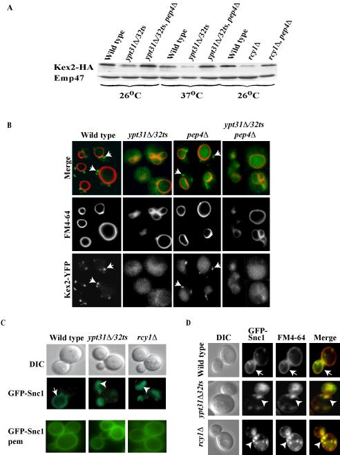
Ypt31/32 and Rcy1 play a role in the recycling of the Kex2 and Snc1 proteins. (A) Ypt31/32 and Rcy1 affect Kex2 stability. Wild-type (NSY125), ypt31Δ/32ts (NSY348), ypt31Δ/32ts pep4Δ (NSY355), rcy1Δ (NSY818), and rcy1Δ pep4Δ (NSY819) cells expressing HA-tagged Kex2 protein were grown entirely at 26°C or shifted to 37°C for 90 min before harvest as indicated. Cell lysates were prepared and Kex2 protein level was measured by Western blot analysis by using anti-HA antibodies. Total protein determination and Emp47 were used as loading and blotting controls. Typical results representing five experiments are shown. (B) Microscopic assay showing that Ypt31/32 GTPases are important for the recycling of Kex2 to the Golgi. Wild-type (NSY970), pep4Δ (NSY971), ypt31Δ/32ts (NSY972), and ypt31Δ/32ts pep4Δ (NSY973) cells expressing endogenous Kex2 tagged with YFP on the chromosome were grown at 26°C. FM4-64 was internalized for 30 min to mark the vacuolar membrane. Intracellular localization was determined by direct fluorescence using an FITC filter for Kex2-YFP and Texas Red filter for FM4-64. Kex2-YFP in the Golgi is seen as green dots outside the vacuolar ring (arrows) in wild-type but not mutant cells. (C) Ypt31/32 and Rcy1 are involved in Snc1 recycling to the plasma membrane. Wild-type (NSY729), ypt31Δ/32ts (NSY733), and rcy1Δ (NSY737) cells expressing GFP-tagged Snc1 protein were grown at 26°C. GFP-Snc1 intracellular localization was determined by direct fluorescence by using an FITC filter. GFP-Snc1-pem, which cannot be internalized, is shown at the bottom. This form of Snc1 localizes to the PM in wild-type and mutant cells. (D) Snc1 localizes to early endosomes in ypt31Δ/32ts and rcy1Δ mutant cells. Cells were grown and treated as in C, except that early endosomes were marked by FM4-64 internalized for 5 min (some FM4-64 is still on the plasma membrane after such a short pulse). In C and D, arrows indicate PM staining, and arrowheads point to internalized GFP-Snc1; the contour of the cells is shown in the DIC image.

