Figure 4.
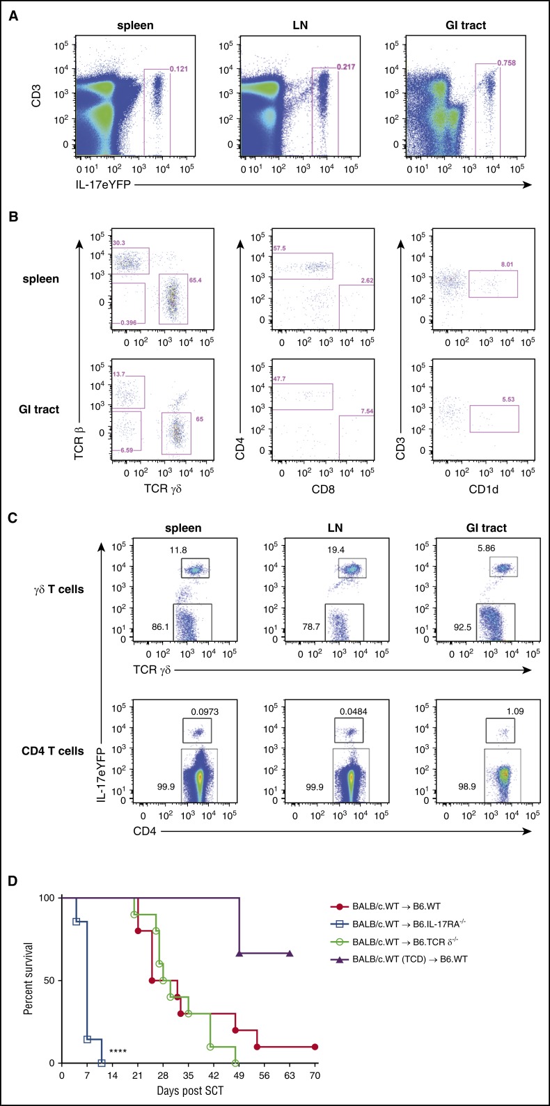
Recipient γδ T cells and conventional CD4 T cells are the predominant source of IL-17A after SCT. (A) Representative plots showing the frequency of IL-17eYFP+ cells in spleen, LN, and the GI tract of naïve B6.IL-17eYFP reporter mice. (B) Phenotype of the IL-17eYFP+ cells in the spleen and GI tract; γδ T cells (TCRγδ+), conventional CD4 T cells (TCRβ+ CD4+), conventional CD8 T cells (TCRβ+ CD8+), and NKT cells (CD3+ CD1d+) are shown. (C) Proportion of IL-17eYFP+ vs IL-17eYFPneg γδ T cells and conventional CD4 T cells in spleen, LN, and the GI tract of naïve B6.IL-17eYFP reporter mice is shown. Data combined from individual mice (n = 3) for all tissues except the GI tract where the mice were pooled. (D) Lethally irradiated B6.WT (red filled circles), B6.IL-17RA−/− (blue open squares), or B6.TCRδ−/− (green open circles) mice received either T-cell replete or TCD grafts (purple filled triangles) (n = 7 to 10 per group; TCD group, n =3) from G-CSF immobilized BALB/c.WT donors. Survival is represented by Kaplan-Meier analysis. Data combined from 2 replicate experiments are shown. ****P < .0001, WT vs IL-17RA−/− recipients. LN, lymph node.

