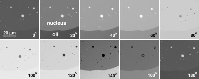
Figure 2.
Small portion of a GV isolated in oil. A Xenopus GV was isolated and gently squashed in oil of refractive index = 1.3512. It was then observed in the interferometer microscope. A small part of the nucleus, which contains nucleoli and speckles, occupies the upper portion of the images. The boundary between the nucleus and oil runs horizontally across the lower part of the images. Charge-coupled device images were taken at 20° intervals, as the compensator was rotated through 180° (= 1 wavelength of retardation). Quantitative analysis showed that in this sample the nucleoplasm was retarded by 3.8° relative to the oil. That is, the intensity of the nucleoplasm at n° was equal to the intensity of the oil at (n + 3.8)°. When a GV is mounted in oil of refractive index = 1.3544, the nucleoplasm and oil have identical intensities at all settings of the compensator.

