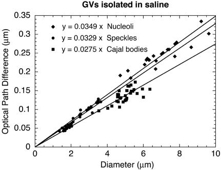
Figure 6.
OPD as a function of organelle diameter (organelles in saline). GVs were isolated in a saline solution (isolation medium), the nuclear envelope was removed, and organelles were centrifuged onto a glass slide. The OPDs of organelles were measured relative to the surrounding saline solution. As in Figure 5, OPDs were plotted as a function of organelle diameter, and the data were fitted to a straight line passing through the origin. Refractive indices estimated for these isolated organelles are similar to those measured within intact GVs.

