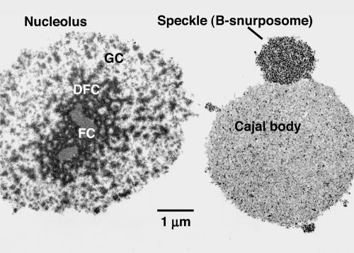
Figure 8.
Electron micrograph of organelles from the GV. Organelles from a Xenopus GV were centrifuged onto a microscope slide, fixed, embedded, and sectioned for electron microscopy. The thin section shows that there are no membranes around the three major organelles: nucleoli, speckles, and CBs. Thus, the interior of the organelles seems to be accessible to the external medium. Injection experiments show that dextran does, in fact, penetrate the organelles in unfixed GVs (Figure 9). FC, DFC, and GC refer to the fibrillar center, dense fibrillar component, and granular component of the nucleolus, respectively.

