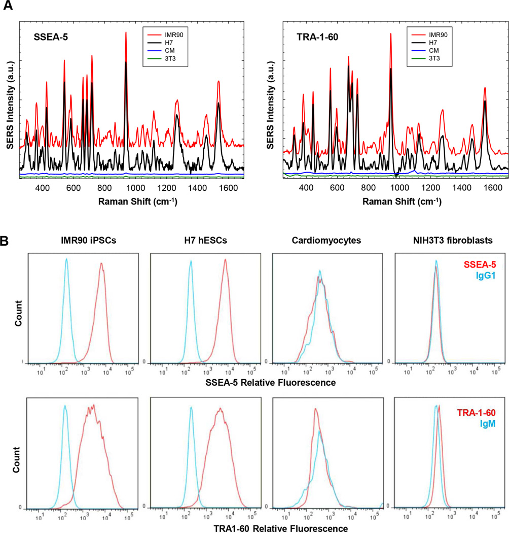
Figure 4.
Specificity of SSEA-5-conjugated and TRA-1-60-conjugated nanoparticles for the detection of hPSCs. (A) Cells (500,000 cells/test) were pretreated with an endogenous biotin-blocking reagent and then incubated with nanoparticles for 30 min. Following washing, the cells were centrifuged and the cell pellet was analyzed using a Raman system for each ensemble measurement. Excitation wavelength: 785 nm; laser power: 70 mW; integration time: 1 sec. Note: SERS signals from SSEA-5-conjugated and TRA-1-60-conjugated nanoparticles were detected in pluripotent stem cells, IMR-90 iPSCs (red) and H7 hESCs (black), but not in non-stem cells, primary rat cardiomyocytes (CM, blue) or NIH3T3 fibroblasts (3T3, green). (B) Flow cytometry analysis of pluripotent stem cell surface markers. SSEA-5+ cells and TRA-1-60+ cells were detected in IMR-90 iPSCs and H7 hESCs, but not in cardiomyocytes or NIH3T3 fibroblasts.

