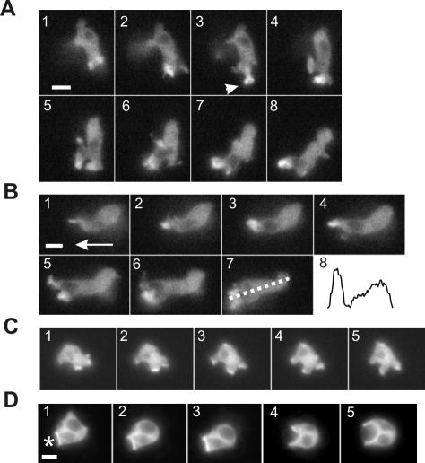
Figure 5.
PakB-GFP localizes to the leading edge of migrating cells. Time-lapse fluorescent images of live PakB-null cells expressing PakB-GFP. Also see Supplemental Movie S1 available at http://www.molbiolcell.org. (A) Images taken at 10-s intervals of a 6-h starved amoeba that traveled from top to bottom (1-4) and then changed direction to migrate from right to left (5-8). PakB-GFP redistributes from the original leading edge to the new leading edge and in 3, momentarily shows up in a cup-shaped structure (arrow) at the tip of the extended pseudopod. (B) Images taken at 10-s intervals of the same cell at a later time migrating in the direction shown by the arrow in 1. Panel 8 shows the fluorescence intensity from the leading edge to the posterior of the cell measured as indicated by the dotted line in 7. (C) Images taken at 20-s intervals of a 2-h starved amoeba taking up fluids by macropinocytosis. (D) Images taken at 20-s intervals of a 2-h starved amoeba ingesting yeast particles. The asterisk in 1 shows the location of the yeast particle. Bars, 5 μm.

