Figure 7.
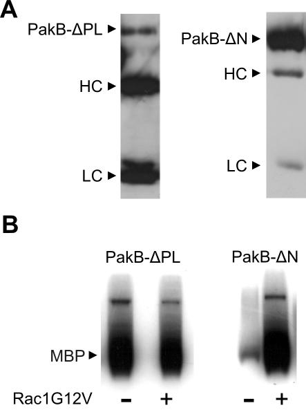
PakB-ΔPL exhibits constitutive kinase activity. (A) Immunoblot analysis of FLAG-PakB-ΔPL (left) and FLAG-PakB-ΔN (right) immunoprecipitated from 2.5 × 10⁷ cells by using anti-FLAG M2 agarose beads and probed with anti-FLAG M5 antibody. Immunoprecipitates were resuspended in 50 μl of 1× SDS sample buffer, and aliquots of 25 and 5 μl were loaded on the SDS gel for the PakB-ΔPL and PakB-ΔN samples, respectively. HC and LC refer to the antibody heavy chain and light chain, respectively. (B) In vitro kinase assays were performed using a 10-μl aliquot of the FLAG-PakB-ΔPL or FLAG-PakB-ΔN immunoprecipitate in the presence or absence of human Rac1G12V with myelin basic protein (MBP) as substrate. The autoradiograph shows the incorporation of 32P into MBP after a 60-min incubation.

