
Figure 4.
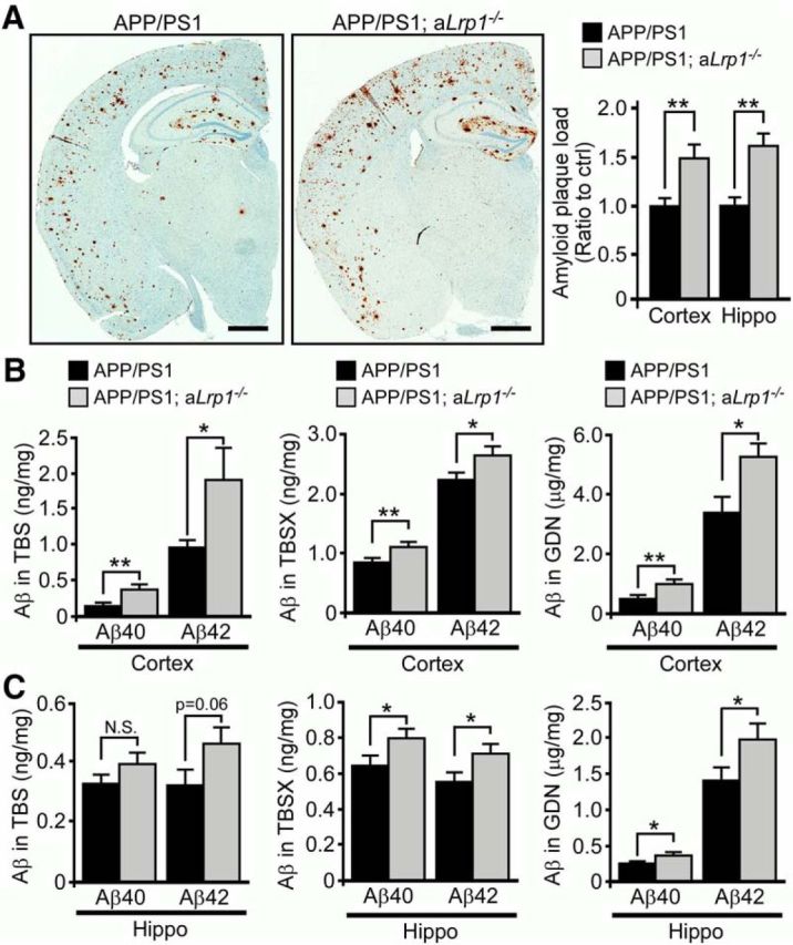
LRP1 deficiency in astrocytes enhances amyloid deposition in APP/PS1 mice. A, Brain sections obtained from 12-month-old APP/PS1 and APP/PS1; aLrp1−/− mice were immunostained for Aβ. Scale bar, 1 mm. The percentage of area covered by amyloid plaques was quantified. B, C, Cortical and hippocampal brain tissues were fractionated into TBS-soluble, detergent-soluble (TBSX), and insoluble (guanidine-HCl, GDN) fractions. Aβ levels from each fraction were quantified by ELISA. Data are mean ± SEM (n = 9 or 10/group). *p < 0.05 (two-tailed Student's t test). **p < 0.01 (two-tailed Student's t test).