Figure 1.
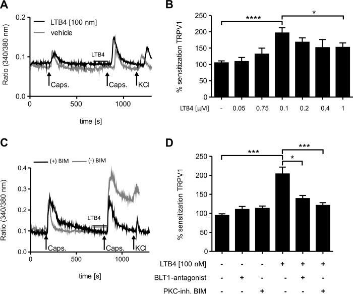
LTB4 sensitizes TRPV1-mediated calcium influx through BLT1. A–D, calcium imaging from DRG culture from C57BL/6N (A and B) or BLT1 knock-out mice (C and D). A, capsaicin (200 nm) stimulates intracellular calcium increases in DRG neurons (black line). Incubation with LTB4 before a second stimulation sensitizes intracellular calcium increases (gray line). Neurons were identified by responses to KCl (50 mm). Shown is a representative trace. B, same as A except that different LTB4 concentrations were used. Sensitization represents the ratio of the second peak by the first peak. The data are presented as means ± S.E. (n = 32–86; one-way ANOVA; Kruskal-Wallis test; Dunn's post hoc test). *, p < 0.05; ****, p < 0.0001. C and D, same as A and B except that DRGs were incubated with the BLT1 antagonist U75302 (1 μm) or the PKC inhibitor BIM (1 μm) before LTB4 treatment. The data are shown as means ± S.E. (n = 45–118). DRGs of three animals were evaluated. Statistics were derived using one-way ANOVA (Kruskal-Wallis test, Dunn's post hoc test). *, p < 0.05; ***, p < 0.001.

