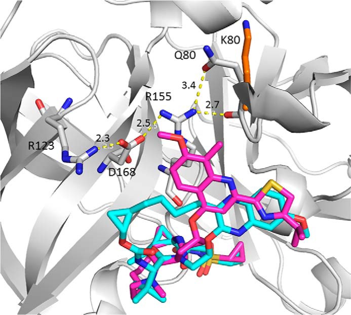
Figure 2.

Superposition of the crystal structures of grazoprevir (cyan) and simeprevir (magenta) bound to HCV NS3/4A protease (PDB codes 3SUD and 3KEE). The protein is represented as ribbon, and only residues Arg-123, Asp-168, Arg-155, and Gln-80/Lys-80 that form the salt bridge/hydrogen bonding network are shown (gray, carbon; red, oxygen; blue, nitrogen). The Lys-80 side chain is also highlighted (orange, carbon). The dashed yellow lines represent the distance in angstroms as labeled.
