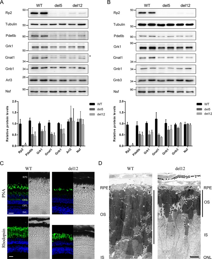
Figure 2.
The del12 mutant zebrafish showed decreased rod phototransduction proteins and progressive retinal degeneration. A and B, protein levels of the rod phototransduction proteins (Pde6b, Grk1, Gnat1, and Gnb1) and cone phototransduction protein Gnb3 as well as the two RP2-interacting protein Arl3 and Nsf were detected by Western blot in 1-month-old (A) and 2-month-old (B) WT, del5, and del12 mutant zebrafish. Tubulin was used as a loading control. The asterisk in A indicates a nonspecific bands produced by the anti-GNAT1 antibody. The quantitative results are shown as the mean with S.D. (n = 6) in the lower panels, respectively. C, retinal cryosections from WT and del12 mutant zebrafish were stained with PNA (upper panel) and the anti-rhodopsin (4D2) antibody (lower panel) to label the outer segments of cones and rods at the age of 6 months. Reduction in the thickness of the rod outer segment layer is seen in del12 mutant zebrafish. Scale bars: 20 μm. D, retinal ultrastructure analysis of 7-month-old WT and del12 mutant zebrafish revealed significantly shortened outer segments of photoreceptors in del12 mutant zebrafish. RPE, retinal pigment epithelium; OS, outer segment; IS, inner segment; ONL, outer nuclear layer. Scale bars: 5 μm.

