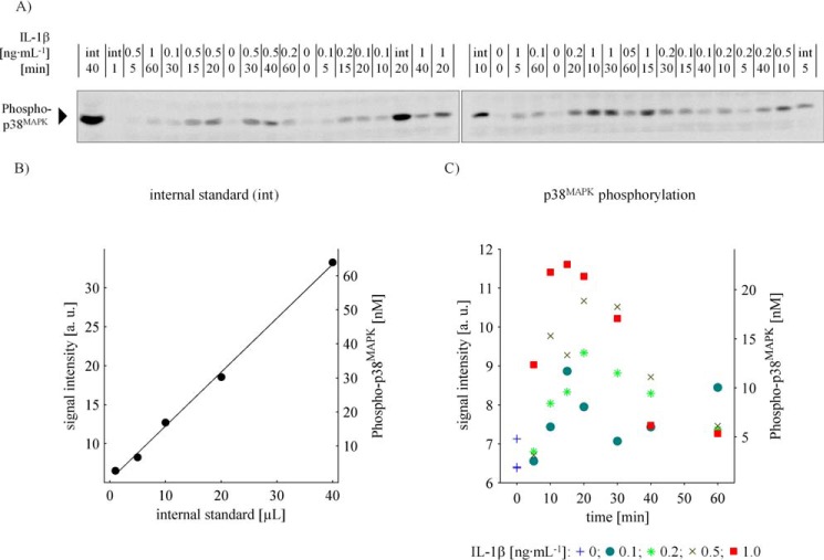
Figure 1.
Quantification of p38MAPK phosphorylation in primary mouse hepatocytes. A, immunoblotting of whole cell lysates in co-separation with an internal standard affords the quantification of intracellular phosphorylated p38MAPK molecule concentration. Primary mouse hepatocytes were treated with 0, 0.1, 0.5, and 1 ng·ml−1 IL-1β, respectively, for up to 60 min. The cells lysed in Triton lysis buffer and specimen were applied randomly to SDS-PAGE and subsequent Western blotting in co-separation with a dilution series (1, 5, 10, 20, and 40 μl) of an internal standard (int). After incubation with specific antibodies, the nitrocellulose membrane was cut in half to fit on the documentation screen, and chemiluminescence was captured at a 16-bit charge-coupled device. B and C, quantitative determination of IL-1β-induced p38MAPK phosphorylation kinetics. The intensity of the chemiluminescent signals was quantified, and, in consideration of cell volume and the internal standard, the intracellular concentration of p38MAPK phosphorylation as a function of IL-1β and time was quantified. a. u.: arbitrary units.

