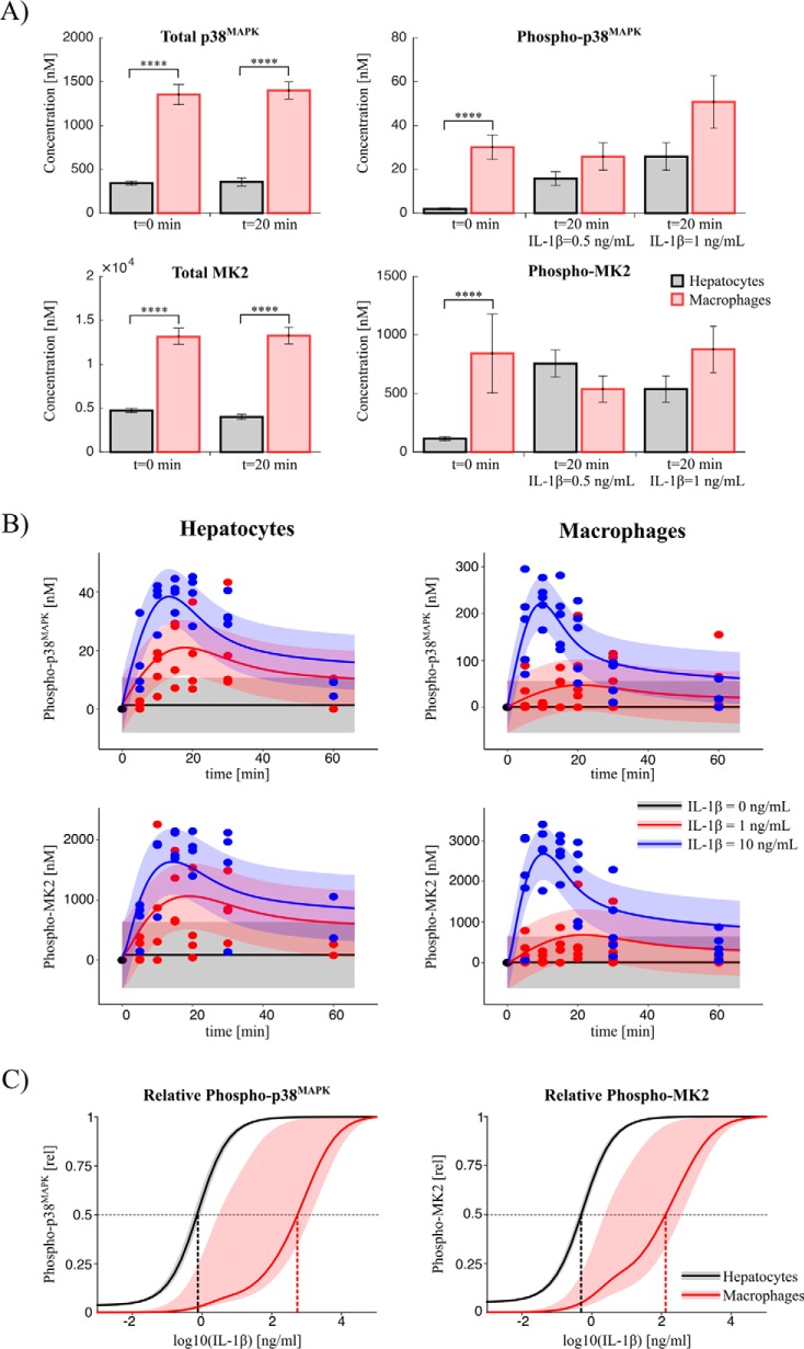
Figure 9.
Comparative analyses between hepatocytes and macrophages. A, absolute concentrations. The concentrations of total and phosphorylated p38MAPK and MK2 were measured with the internal standard for different durations and concentrations of IL-1β treatment of macrophages (red) and hepatocytes (gray), respectively. The total concentrations are significantly higher in macrophages in comparison with hepatocytes. The criteria for significance were a two-way analysis of variance and post hoc pairwise comparisons of each IL-1β treatment condition of hepatocytes and macrophages, respectively, based on multiple two-tailed t tests. The p values were adjusted according to Bonferroni to account for the multiple comparisons problem. ****, p < 0.0001. B, kinetics. The kinetics of phosphorylated MK2 and p38MAPK were measured in macrophages and hepatocytes for different IL-1β inputs (points). For hepatocytes, the solid lines indicate the prediction of the calibrated model. For macrophages, the mathematical model was fitted to the measured macrophage data (solid line). C, model prediction of dose responses. Shown is relative phosphorylation dependent on the IL-1β input dose as predicted with the calibrated models. The bands show the prediction uncertainty calculated by integrating all parameter sets along the corresponding profile likelihoods.

