
Figure 5.
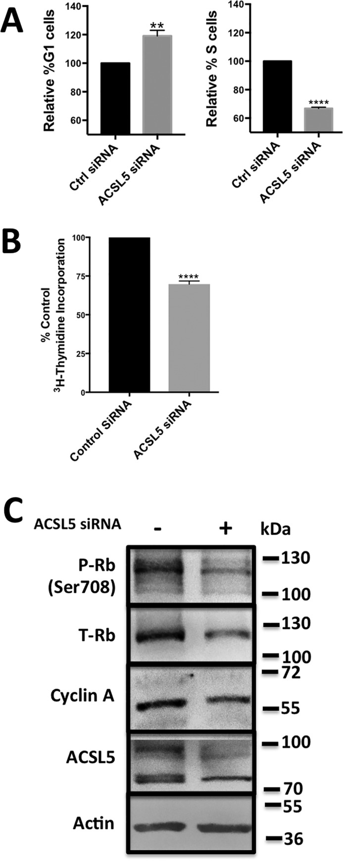
Suppressing ACSL5 expression causes G1 cell cycle arrest. A, Calu-1 cells were plated at 40% confluence and transfected with ACSL5 siRNA (25 nm) or control (Ctrl) siRNA. 96 h post-transfection, cells were collected and analyzed by flow cytometry. Data are shown in terms of the percentage of change in the number of cells in G1 and S phases of the cell cycle. Mean values for G1 phase cells were 53.4% ± 10.4% and 25.2% ± 9.4% for S phase cells in the controls and were normalized to 100%. Analysis was performed on at least 10,000 cells for each condition. B, Calu-1 cells were plated at 40% confluence and transfected with ACSL5 siRNA or control siRNA as in A. 48 h post-transfection, [3H]thymidine was added. After 24 h, cells were collected, and total radioactivity was determined by scintillation counter. Statistical significance (p values) for A and B were determined by Student's two-tailed unpaired t test. **, p ≤ 0.01; ****, p ≤ 0.0001 compared with the control. C, Calu-1 cells were plated at 40% confluency and transfected with ACSl5 siRNA (25 nm) or control siRNA. 96 h post-transfection, cells were collected and analyzed for the levels of ACSL5, phospho-Rb (P-Rb), total Rb (T-Rb), cyclin A, and actin by Western blotting. The Western blot shown is representative of experiments repeated twice.