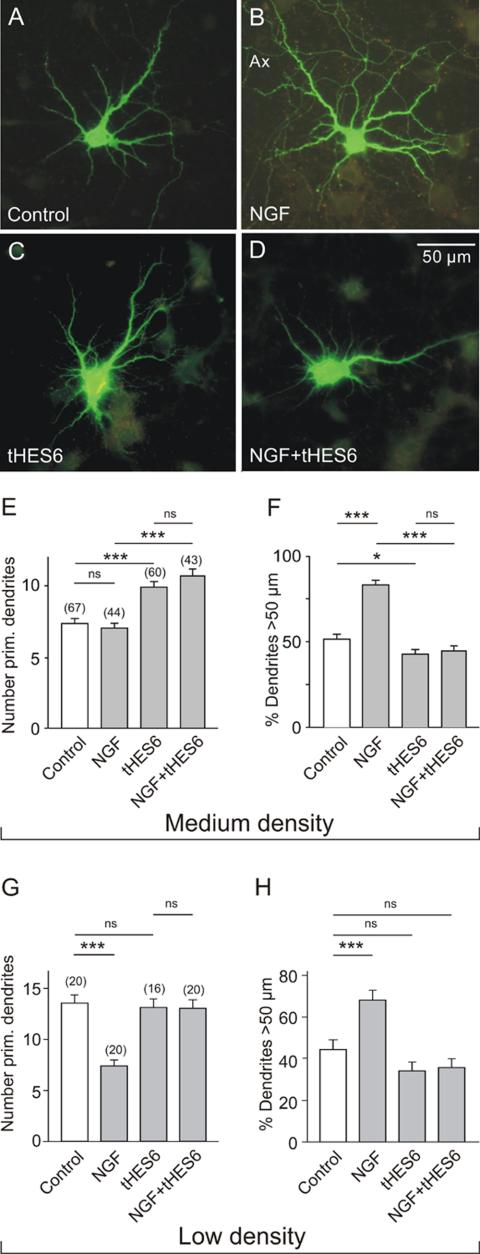
Figure 6.
Effect of Hes6 expression on the dendrite morphology of hippocampal neurons in medium- and low-density cultures. (A-D) Digital fluorescence images of EGFP-labeled neurons in medium-density cultures after 16 h in control medium (A and C) or in medium with NGF (100 ng/ml; B and D). E17 hippocampal neurons were cultured for 7 DIV and then transfected with pIRES vectors that either induced expression of EGFP alone (A and B) or the expression of EGFP and Hes6 (C and D). Note the larger dendritic field of the NGF-treated neuron (B) and the lack of response to NGF in the cell expressing Hes6 (D). (E and F) Results of morphometric evaluation of medium density cultures (50,000 cells/cm2). Note that NGF was unable to prevent the Hes6-stimulated outgrowth of new primary dendrites. Hes6-transfection also effectively blocked the NGF-induced dendrite elongation. (G and H) Results of morphometric evaluation of low cell density cultures (15,000 cells/cm2). Note that NGF decreased the number of primary dendrites and increased the relative length of dendrites, but these effects were prevented by transfection with Hes6. Symbols and parameters as in Figure 2.

