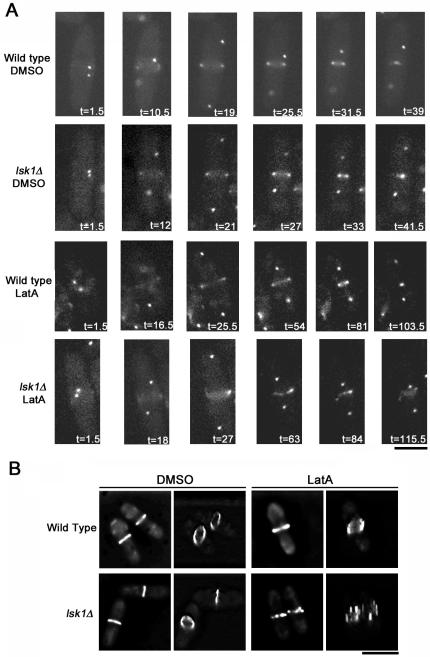
Figure 4.
Actomyosin rings fragment in lsk1Δ mutant backgrounds in the presence of 0.3 μM LatA. (A) Time-lapse microscopy of wild-type (Supplementary Time-Lapse Videos 5 and 7) and lsk1Δ cells (Supplementary Time-Lapse Videos 6 and 8) carrying an integrated copy of both Rlc1-GFP and Sid4-GFP. Cells were treated with DMSO (Supplementary Time-Lapse Videos 5 and 6) or 0.3 μM LatA (Supplementary Time-Lapse Videos 7 and 8) before being imaged at 90-s intervals. Bar, 5 μm. (B) Maximum projections (left-hand panels) and tilted 3D reconstructions (right-hand panels) of deconvolved Z-stacks of wild-type and lsk1Δ cells expressing rlc1-GFP treated with 0.3 μM LatA, or DMSO, for 30 min. Bar, 10 μm.

