Figure 2.
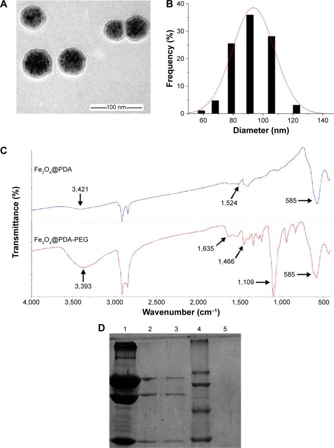
Confirmation of the bioconjugation of EGFR antibody to Fe3O4@PDA NPs.
Notes: (A) TEM image and (B) size distribution histogram of the Fe3O4@PDA NPs. (C) FT-IR spectra of Fe3O4@PDA and Fe3O4@PDA-PEG. (D) SDS-PAGE analysis of Fe3O4@ PDA-PEG-EGFR and EGFR antibody. The Coomassie blue-stained gel analysis revealed the successful cross-linking of EGFR antibody molecules on the surface of the Fe3O4@PDA-PEG NPs. Lane 1, EGFR antibody; lanes 2 and 3, the bioconjugated Fe3O4@PDA-PEG-EGFR NPs; lane 4, protein molecular weight marker; lane 5, Fe3O4@PDA-PEG NPs.
Abbreviations: FT-IR, Fourier transform–infrared; SDS-PAGE, sodium dodecyl sulfate–polyacrylamide gel electrophoresis; TEM, transmission electron microscope; PDA, polydopamine; PEG, polyethylene glycol; NP, nanoparticle.

