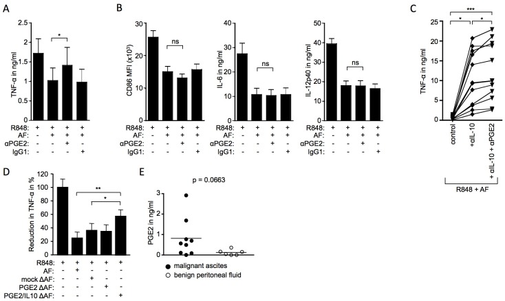
Fig 7. Paracrine OC-associated PGE2 impairs TLR-mediated DC activation and distinguishes malignant carcinoma from benign ovarian conditions.
Monocyte-derived DC were stimulated overnight with 3μg/ml R848. (A-B) Malignant ascites and neutralizing antibodies (5μg/ml) against PGE2 were added as indicated; n = 11 (11 independent experiments; monocyte-derived DC from 4 different healthy donors, cultured with 2 (n = 1), or 3 (n = 3) different ascites samples) (C) Neutralizing antibodies against IL-10 and PGE2 were added as indicated to cultures containing 3μg/ml R848 and 10% ascites; n = 9 (9 independent experiments, monocyte-derived DC from 4 different healthy controls, cultured with 1 (n = 1), 2 (n = 1) or 3 (n = 2) different ascites samples). (D) Malignant ascites which had previously been depleted of PGE2 (PGE2 ΔAF) or PGE2 and IL-10 (PGE2/IL10 ΔAF) or which had undergone mock depletion (mock ΔAF) was added as indicated. As control, DC cultures were treated with R848 and untreated ascites (AF); n = 4 (4 independent experiments; monocyte-derived DC from 1 healthy donor, cultured with ascites samples from two individual ovarian carcinoma patients). The level of TNFα is expressed in relation to the TNFα levels induced in response to LPS, which were set to 100%. (A-D) Cytokine levels in cell culture supernatants were measured by sandwich ELISA. One-way ANOVA (Friedman test with Dunn post test); * = p<0.05; ** = p<0.01; *** = p<0.001; ns = not significant. (E) PGE2 levels in malignant ascites (n = 9) and benign peritoneal fluid (n = 6) were measured by sandwich ELISA. Mann-Whitney U test was used for statistical analysis.

