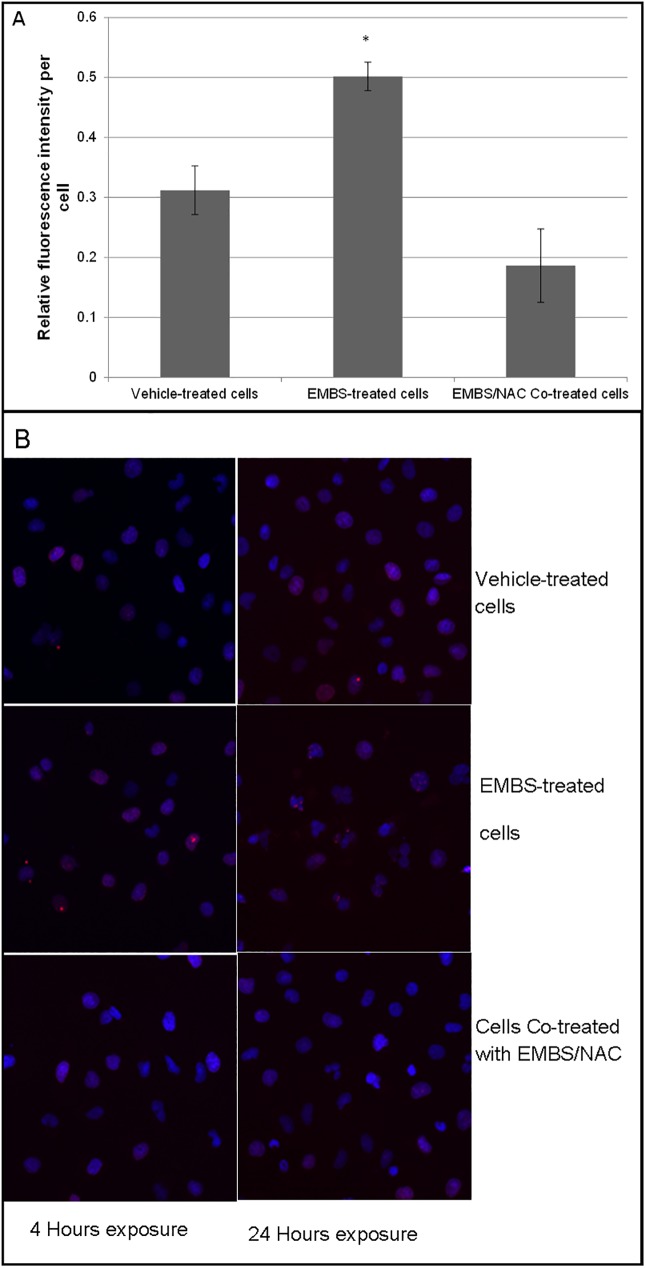
Fig 5. EMBS induces DNA double strand breaks and endoreduplication.
(A) Confocal images of cells treated for 4 h with 0.4 μM EMBS or EMBS and 20 mM NAC were quantified by determining pixel values for H2A staining divided by the number of cells per image. The average of 100 cells per treatment was used and the average of three independent experiments are represented with s.e.m. represented by the error bars. A significant difference between the DMSO treated and EMBS treated cells was observed at a P-value of 0.25. (B) Cells incubated with EMBS or EMBS and NAC for 24 h were stained for phosphorylated H2A (red) and DAPI (blue). Representative images show an increase in the number of deformed nuclei in EMBS treated cells, while these were mostly absent in control or NAC-treated cells.

