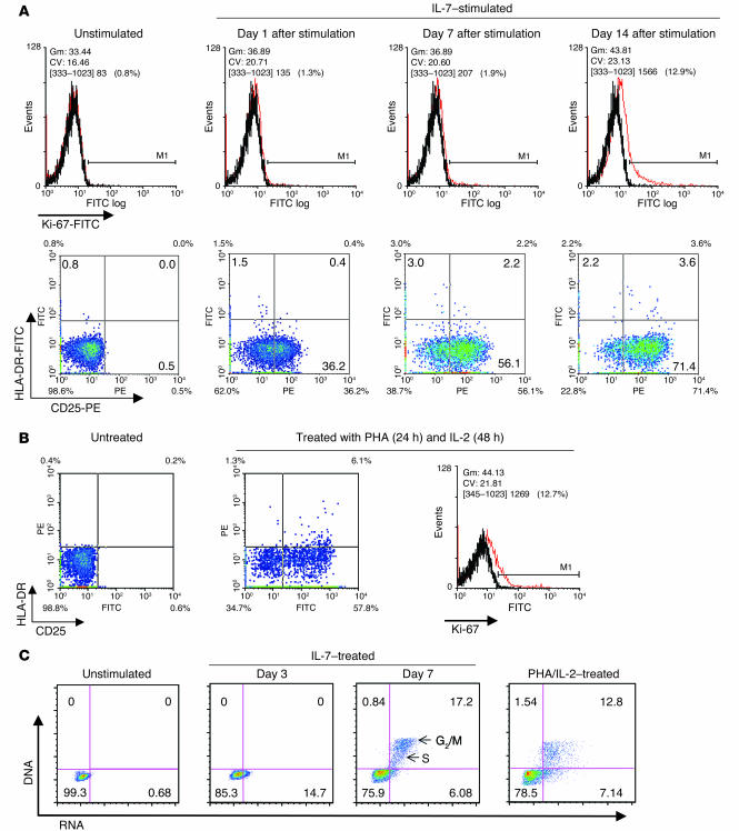
Figure 2.
Stimulatory patterns of resting CD4+ T lymphocytes by IL-7. (A and B) Detection of cell activation markers in IL-7–treated or PHA/IL-2–treated human resting CD4+ T lymphocytes. Freshly isolated resting CD4+ T lymphocytes stimulated with IL-7 or PHA/IL-2 were stained with FITC-conjugated anti–KI-67, anti–HLA-DR, and PE-conjugated anti-CD25. Three independent experiments were performed, of which one representative is illustrated. Gm, geometric mean; CV, coefficient of variance. (C) Effects of IL-7 on the cell cycle of human peripheral blood resting CD4+ T lymphocytes. IL-7 alone or PHA/IL-2–stimulated, initially resting CD4+ T lymphocytes are shown with the cell-cycle status indicated in each quadrant. DNA is depicted on the vertical axis and RNA is shown on the horizontal axis. Positions indicating stages in the cell cycle are illustrated in the third panel. The cell cultures either alone or in the presence of IL-7 were continued for a full 7 days. PHA/IL-2–stimulated cells were treated with PHA (5 μg/ml) for 48 hours and then IL-2 (10 ng/ml) for 3 days.

