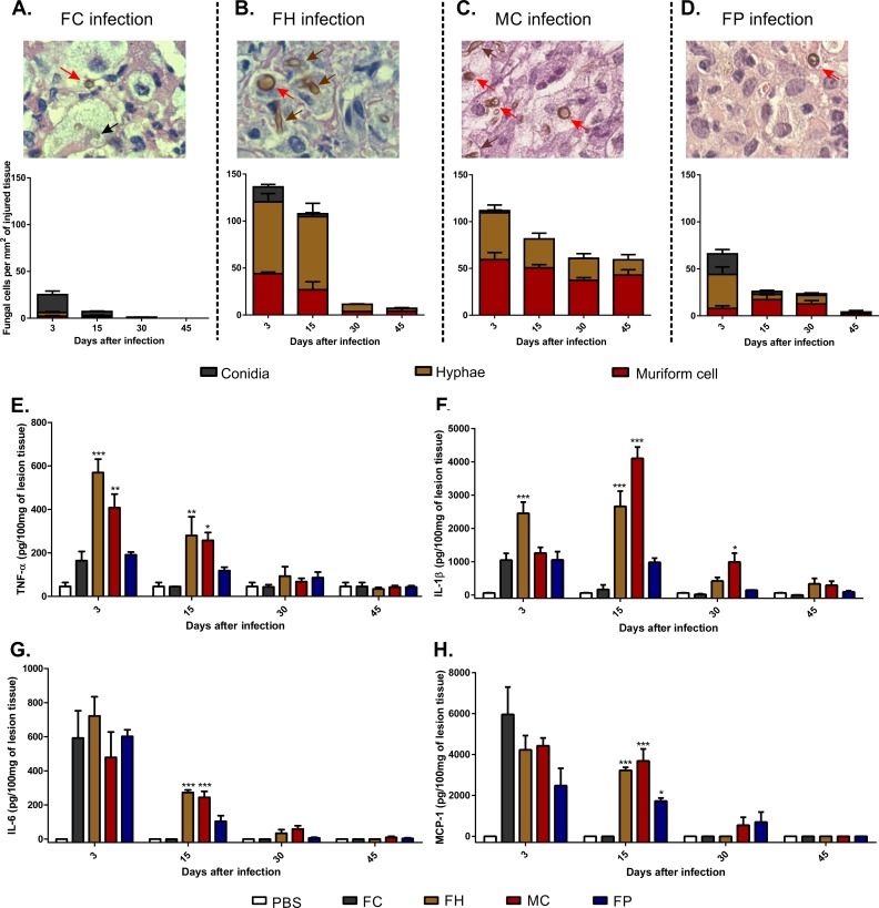
Fig 2. Quantification of F. pedrosoi fungal cells in the tissue and production of pro-inflammatory cytokines in the course of murine CBM.
Fungal cells were counted on twenty fields chosen at random in histopathological slides with the aid of a counting reticle (A-D). In all groups muriform cells (red arrows) could be identified after 15 days after infection. At the same time, hyphal fragments (brown arrow) were also present in animals infected either with hyphae (FH) (B) or muriform cells (MC) (C). Few conidia (black arrow) are observed after 15 days of infection in animals infected with F. pedrosoi conidia (FC) (A). Cell counts were expressed as cells per mm2 of injured tissue. 1000x magnification (A-D). After 15 days of infection, high levels of TNF-α (E), IL-1β (F), IL-6 (G) and MCP-1 (H) were observed in groups with higher numbers of hyphae and muriform cells in the tissue. Cytokine production was measured by ELISA from homogenized footpad tissue. *P<0.05, **P<0.01 and ***P<0.001 compared to FC group.

