Figure 9.
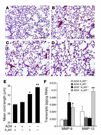
Alveolar destruction in ADA/A1AR double-knockout mice. ADA–/– mice were treated with ADA enzyme therapy from birth until postnatal day 14 as described in Methods. Lungs from ADA–/– mice and age-matched ADA+ mice were collected 14 days after the cessation of ADA enzyme therapy and processed for H&E staining. Images show (A) lung from an ADA+A1AR+/+ mouse, (B) lung from an ADA+A1AR–/– mouse, (C) lung from an ADA–/–A1AR+/+ mouse, and (D) lung from an ADA–/–A1AR–/– mouse. Images are representative of 10 animals from each group. Scale bar: 100 μm. (E) Alveolar airway sizes were calculated using Image-Pro Plus (Media Cybernetics). Data are presented as mean cord length ± SEM; n = 5. (F) Levels of MMP-9 and MMP-12 transcripts were determined in whole-lung extracts from postnatal day 14 mice using quantitative RT-PCR. Data are presented as mean pg of transcripts/μg RNA ± SEM; n = 8 for each. *P – 0.05 compared to ADA+ mice; **P – 0.05 compared to ADA–/–A1AR+/+ mice.

