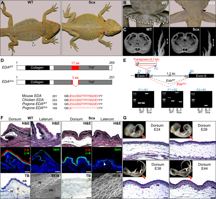
Fig. 3. Characterization of mutant scaleless P. vitticeps lizards.

(A) Dorsal views of adult wild-type (WT) and scaleless (Sca) P. vitticeps lizards. The white arrowhead indicates the presence of large lateral spines in the WT. (B) Ventral views of WT and Sca adult males showing the absence of femoral pores (arrowheads) in mutant lizards. (C) Micro x-ray computed tomography scan virtual sections of the skull (left) and magnified views of the autopod (right) of WT and Sca dragons at birth. White frames indicate the position of the pleurodont regenerating teeth, and double-headed arrows show the relative sizes of claws. (D) Diagram of WT (EDAWT) and mutant scaleless (EDASca) active EDA proteins. The conserved collagen and TNF domains are shown as black and gray boxes, respectively. The most conserved TNF motif [17 amino acids (aa) in WT] is shown in red. The mutant EDA protein has an in-frame deletion of 14 amino acids, as shown by the alignment of EDA protein sequences from mouse, chicken, and WT and Sca P. vitticeps (lower panel). Black numbers represent amino acid position. (E) Upper panel: diagram showing the genomic structure (from exon 7 to 8) of the P. vitticeps Eda gene. Intron length and splice donor (gt) and acceptor (ag) sites are indicated. Blue arrows show the positions of primers used for reverse transcription polymerase chain reaction (RT-PCR) analyses. In the scaleless mutant genome, a transposon of 5.7 kb starting with an alternative splice donor site is inserted in the 3′ end of exon 7, thus leading to an alternative splicing (red dashed lines) of the mutant EdaSca gene in comparison to the splicing of the EdaWT gene (black dashed lines). Lower panels: RT-PCR analysis (g, on genomic DNA; c, on skin cDNA) of WT and Sca animals using the indicated primer combinations. (F) Top row: H&E staining of skin sections from dorsal and lateral body regions of adult WT and scaleless dragons. Middle row: immunofluorescent staining of α-keratins (α-k) and β-keratins (β-k) or laminin (lam; arrowhead shows convoluted basal membrane) in dorsal skin of adult WT and scaleless animals. Bottom row: Toluidine blue (TB) staining of dorsal skin sections and scanning electron microscopy (SEM) images of skin molts from adult WT and scaleless lizards. is, interscale region; os, outer scale region. Scale bars, 50 μm. (G) H&E staining of dorsal skin sections of scaleless P. vitticeps embryos at various developmental stages (indicated as embryonic days after oviposition); red arrows in the top insets indicate the locations of skin sections on lateral views of the corresponding embryos. Scale bars, 100 μm.
