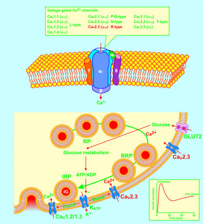
Figure 1.
The functional CaV channel consists of pore-forming subunits CaVα1 and auxiliary subunits CaVβ, CaVα2/δ, and CaVγ. Four types of CaVα1 subunits, designated CaV1.2, CaV1.3, CaV2.1, and CaV2.3, conducting L-, P/Q-, and R-type Ca2+ currents, have been identified in the mouse β cell. Glucose-stimulated insulin secretion is characterized by a rapid first phase of insulin release for about 10 minutes, followed by a nadir, and subsequently a gradually increasing second phase reaching a plateau after 25 to 30 minutes (inset). Insulin-containing granules (IG) are functionally divided into three pools: the reserve pool (RP), the readily releasable pool (RRP), and the immediately releasable pool (IRP). The present consensus is that the KATP channel–dependent mechanisms trigger first-phase insulin secretion from the IRP by opening CaV1.2 and CaV1.3 channels. The KATP channel–independent mechanisms underlie second-phase insulin secretion by recruiting insulin-containing granules from RP and RRP to IRP. The Ca2+ influx through β cell CaV2.3 channels is now demonstrated to play a prominent role in second-phase insulin secretion. CaV1.2/1.3, CaV1.2 channels or CaV1.3 channels; CaV2.3, CaV2.3 channels; Depol, depolarization; GLUT2, glucose transporter 2.

