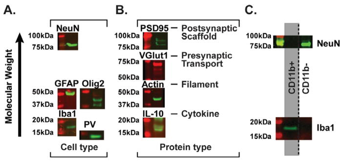
Figure 6. TRIzol-precipitated protein from brain tissue represents multiple cell types and protein classes.
(A) Different cell types inherent in brain tissue are detected in Western blot analysis of precipitated protein. NeuN=neurons, GFAP=astrocytes, Olig2=oligodendrocytes, Iba1=microglia, PV=inhibitory interneurons. (B) Different protein classes are represented in precipitated protein, including synaptic (PSD95, VGlut1), membrane bound (Iba1), nuclear (NeuN), cytoplasmic (actin and ERK1/2 in Fig. 4), and secreted (IL-10) proteins. (C) Protein from specific populations of a single cell suspension (i.e. microglia in the CD11b+ population and other neural cell types in the CD11b− population) is successfully analyzed after solubilization in optimized lysis buffer. The CD11b+ population contains Iba1, but not NeuN protein, while the CD11b− population contains NeuN, but not Iba1 protein.

