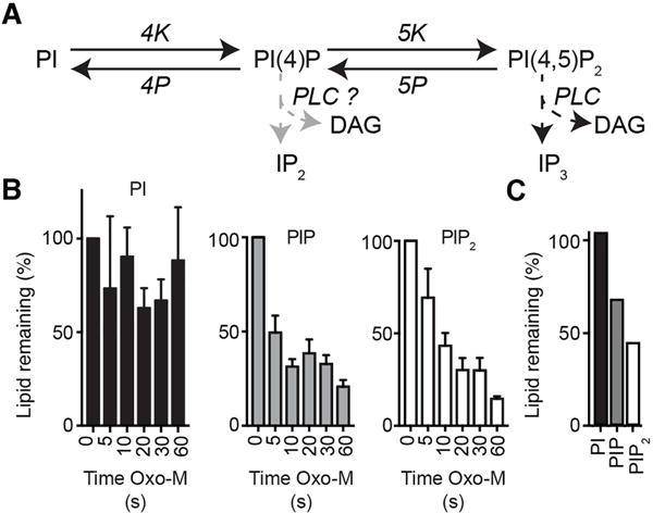
Fig. 5.

UPLC mass spectrometry detects cellular depletion in total PIP and PIP2 pools following activation of phospholipase C (PLC) in CHO-M1 cells. (A) Schematic of cellular phosphoinositide metabolism and the actions of PLC. (B) Summary histograms of the total PI, PIP, and PIP2 signals in control (0 s) and following 5 – 60 s oxotremorine-M (Oxo-M, 10 μM) to activate a stably expressed M1R receptor in the CHO cells (n = 5). Bars are sums of all fatty-acid species in their sodiated and protonated forms. (C) Changes in phosphoinositide lipids of rat pineal glands after stimulation by norepinephrine showing percent remaining in PI, PIP, and PIP2 pools after a 60 min incubation with 1 μM norepinephrine at 37°C compared to untreated control.
