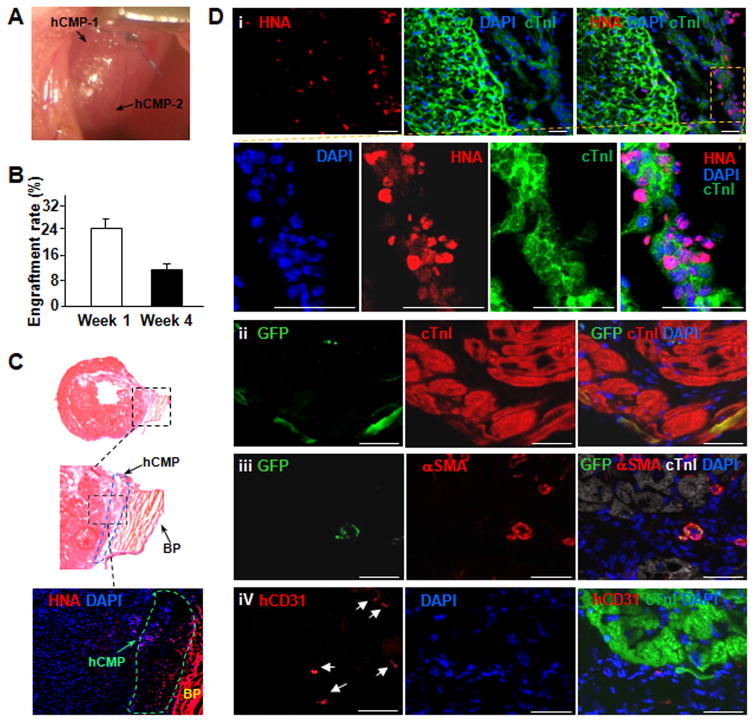
Figure 3. hCMP engraft and survive after transplantation into the hearts of mice with MI.
Myocardial infarction was surgically induced in mice. Animals in the MI+hCMP group were treated with two hCMPs (0.1 million cells total). (A) The representative image showed two transplanted hCMPs on the mouse heart. (B) The engraftment rate was determined in animals from the MI+hCMP group at week 1 and 4 after injury via quantitative PCR measurements of the human Y chromosome (n=4 hearts per time point). (C) The representative HE staining image and human specific nuclear antigen (HNA) immunostaining image showed engrafted hCMP on the epicardial surface of the heart at week 4 after transplantation. BP=bovine pericardium. (D) Sections taken from the region of patch in MI+hCMP animals at week 4 were immufluorescently stained for the presence of HNA, cTnI, green fluorescent protein (GFP), αSMA, and the human isoform of the endothelial marker CD31 (hCD31); nuclei were counterstained with DAPI (scale bar = 50 μm). (i–ii) Engrafted hciPSC-CMs were identified by the expression of both HNA and cTnI (i) or both GFP and cTnI (ii); (iii) engrafted hiPSC-SMCs were identified by the expression of both GFP and αSMA; (iv) engrafted hiPSC-ECs were identified by the expression of hCD31.

