
Fig. 2.
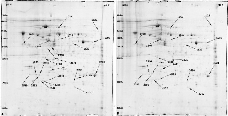
The representative two-dimensional gel electrophoresis images of rCHO-DG44 cultivated in basal SMF as negative control (A) and supplemented with Mix Hydrolysate 8 (B). Differentially expressed protein spots were indicated by arrows.
Official websites use .gov
A
.gov website belongs to an official
government organization in the United States.
Secure .gov websites use HTTPS
A lock (
) or https:// means you've safely
connected to the .gov website. Share sensitive
information only on official, secure websites.
The representative two-dimensional gel electrophoresis images of rCHO-DG44 cultivated in basal SMF as negative control (A) and supplemented with Mix Hydrolysate 8 (B). Differentially expressed protein spots were indicated by arrows.