Fig. 3.

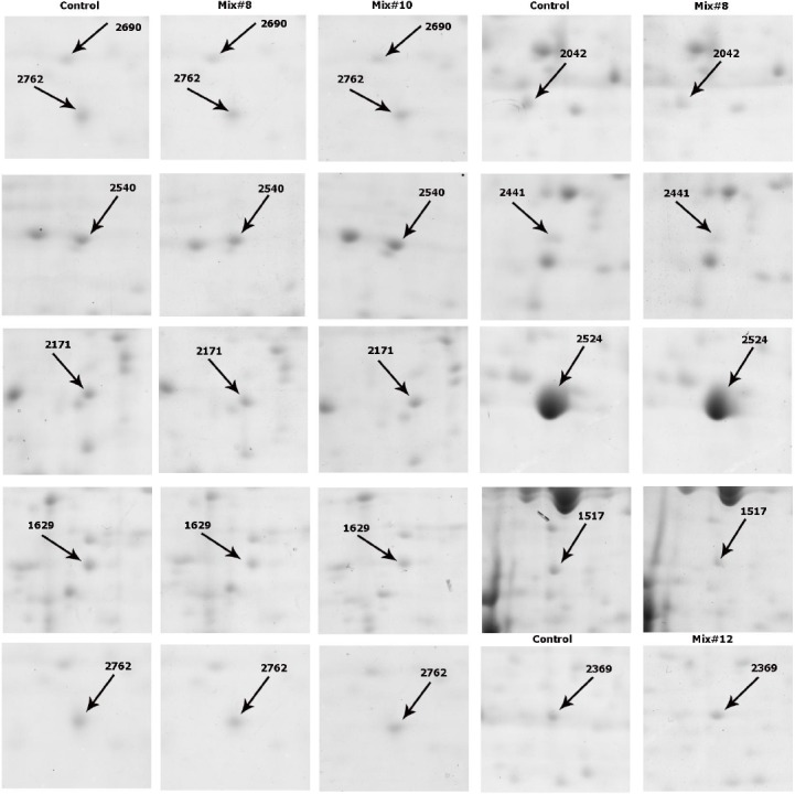
Two dimensional partial images of some differentially expressed protein spots. left panels indicate the gel images of rCHO-DG44 in SFM, and the right panel indicates the gel images of rCHO-DG44 cultivated in Mix. #8, Mix. #10 and Mix. #12.

Two dimensional partial images of some differentially expressed protein spots. left panels indicate the gel images of rCHO-DG44 in SFM, and the right panel indicates the gel images of rCHO-DG44 cultivated in Mix. #8, Mix. #10 and Mix. #12.