
Fig. 3.
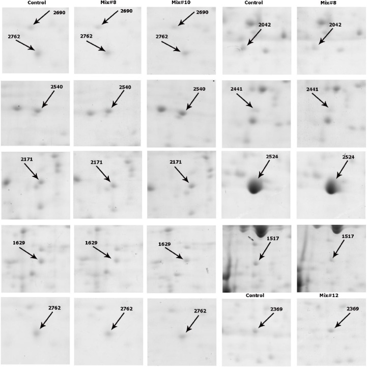
Two dimensional partial images of some differentially expressed protein spots. left panels indicate the gel images of rCHO-DG44 in SFM, and the right panel indicates the gel images of rCHO-DG44 cultivated in Mix. #8, Mix. #10 and Mix. #12.
Official websites use .gov
A
.gov website belongs to an official
government organization in the United States.
Secure .gov websites use HTTPS
A lock (
) or https:// means you've safely
connected to the .gov website. Share sensitive
information only on official, secure websites.
Two dimensional partial images of some differentially expressed protein spots. left panels indicate the gel images of rCHO-DG44 in SFM, and the right panel indicates the gel images of rCHO-DG44 cultivated in Mix. #8, Mix. #10 and Mix. #12.