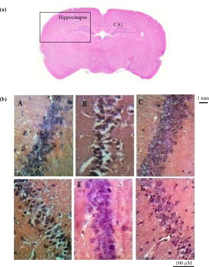
Fig. 2.

Neurodegenerative changes in the hippocampus of pilocarpine-treated rats. (a) coronal section of rat brain in control group; (b) higher magnification of the section seen in part a; Neurodegeneration in CA1 region of hippocampus (20 × objective lens) in control (A, C0; C, C3; E, C5) and pilocarpine-treated rats (B, F0; D, F3; F, F5). Significant neural loss is observed in CA1 area of the hippocampus in F0 group versus control subgroups. F0, after status epilepticus; F3, after development of focal seizures; F5, after acquisition of generalized seizures.
